Showing revision 7.1


-
Translational Cancer Imaging
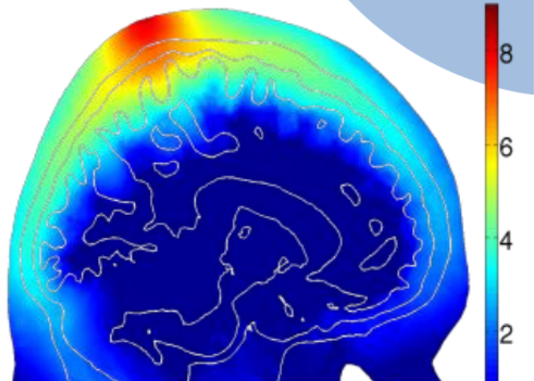
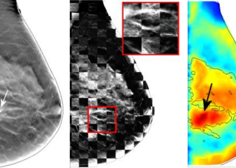
Early diagnosis of breast cancer is critically important. However, the current clinical approach, x-ray mammography, is poor in specificity – over 85% of the recalls yielded benign findings. Furthermore, mammography misses 40% of early stage cancers. Over the past years, our lab has been developing a novel imaging technique to find breast cancers by combining safe, non-invasive near-infrared diffuse optical imaging with high-resolution x-ray mammography. Since 2009, the PI of the lab has been leading this research and conducted a 470-patient clinical study in collaboration with Dr. Daniel Kopans from MGH Avon Center. With our innovative image reconstruction algorithms, we have demonstrated the potentials in differentiating malignant from benign lesions using the functional and structural information together. Our findings were highlighted as a front-cover article in Radiology. In 2011, our lab started a new collaboration with Philips Healthcare to accelerate the clinical translation of this technique. Recently, we published a clinical study showing viability of adding functional breast assessment to all existing mammography systems. In 2016, we filed a provisional patent on a highly effective computer-aided detection (CAD) technique to automatically locate malignant tumors using optical data. These innovations, if successfully translated to the clinic, are expected to positively impact breast cancer patient management by significantly enhancing diagnostic accuracy and early detection. Our breast imaging project was specifically mentioned by the former Massachusetts Governor Deval Patrick in his speech during the “Friends of Cancer Research” forum in 2014.
-
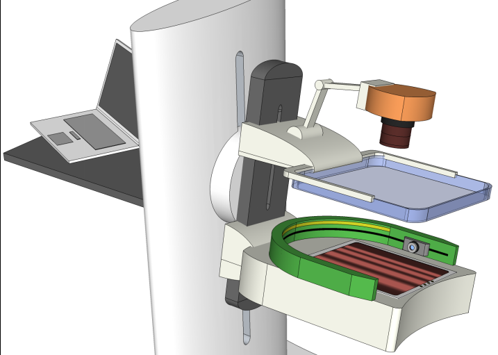 Translational Cancer Imaging
-
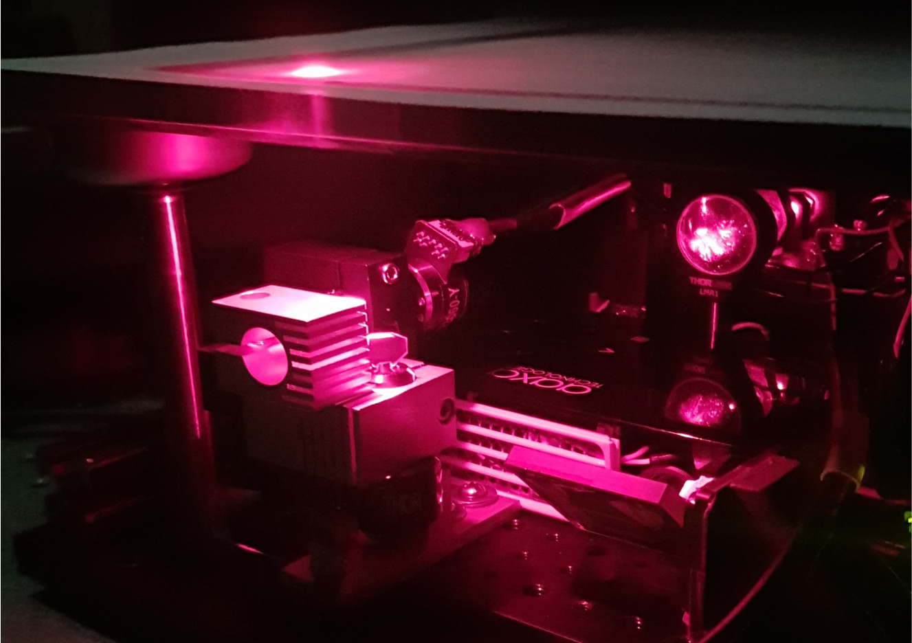
 Biomedical Optics
-
 Monte Carlo eXtreme
-
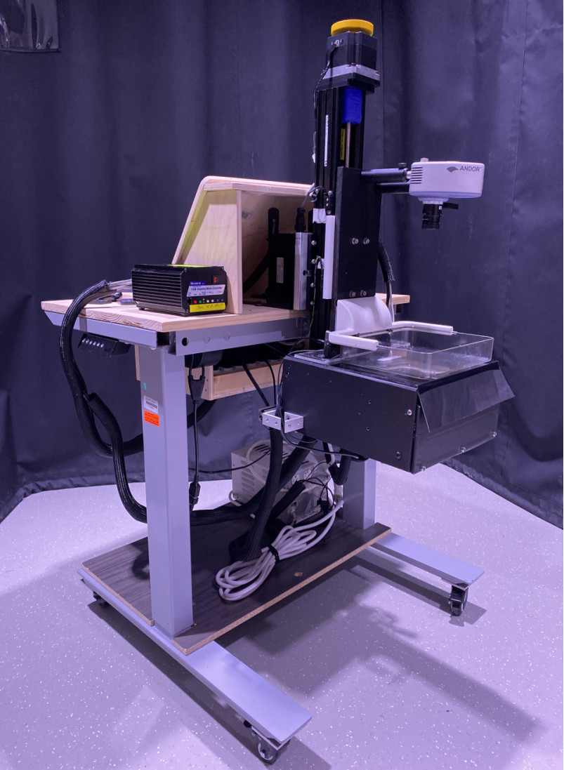
 Point-of-care Devices
-
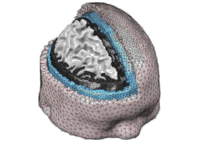
 Multi-modal Imaging