Showing revision 8.14

-
Translational Cancer Imaging
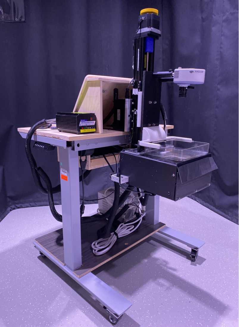
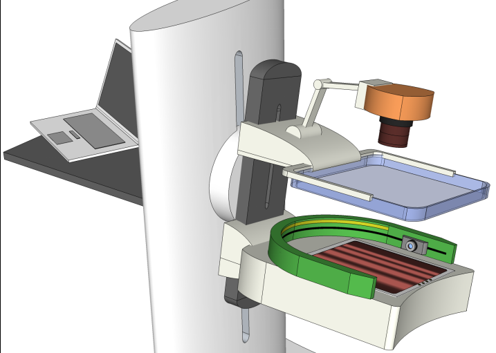
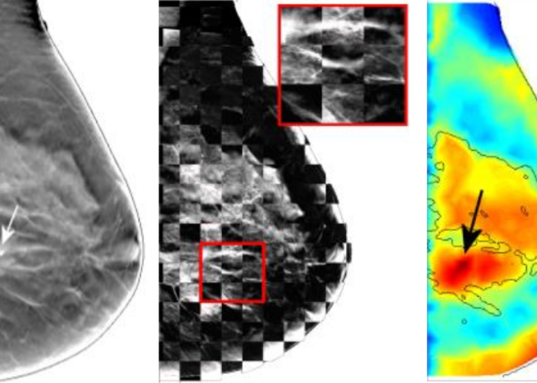
Early diagnosis of breast cancer is critically important. However, the current clinical approach, x-ray mammography, is poor in specificity – over 85% of the recalls yielded benign findings. Furthermore, mammography misses 40% of early stage cancers. Over the past years, our lab has been developing a novel imaging technique to find breast cancers by combining safe, non-invasive near-infrared diffuse optical imaging with high-resolution x-ray mammography. Since 2009, the PI of the lab has been leading this research and conducted a 470-patient clinical study in collaboration with Dr. Daniel Kopans from MGH Avon Center. With our innovative image reconstruction algorithms, we have demonstrated the potentials in differentiating malignant from benign lesions using the functional and structural information together. Our findings were highlighted as a front-cover article in Radiology. In 2011, our lab started a new collaboration with Philips Healthcare to accelerate the clinical translation of this technique. Recently, we published a clinical study showing viability of adding functional breast assessment to all existing mammography systems. In 2016, we filed a provisional patent on a highly effective computer-aided detection (CAD) technique to automatically locate malignant tumors using optical data. These innovations, if successfully translated to the clinic, are expected to positively impact breast cancer patient management by significantly enhancing diagnostic accuracy and early detection. Our breast imaging project was specifically mentioned by the former Massachusetts Governor Deval Patrick in his speech during the “Friends of Cancer Research” forum in 2014. -
Point-of-Care Devices
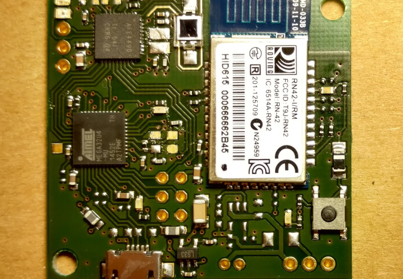
Every year, nearly 3 million newborns die globally within the first 4 weeks of life. In the meantime, over 287,000 women die during pregnancy or following childbirth. Nearly all of these deaths occur in low and middle-income countries (LMICs). We felt the urgency to eliminate this disparity in healthcare and are determined to make innovations to bring life-saving point-of-care diagnostic tools to the resource-poor countries. In 2011, with the funding support from the Bill&Melinda Gates Foundation, we started an investigation on developing low-cost mobile-phone based near-infrared imaging systems. In 2013, we worked with clinical collaborators and initiated another project to develop a mobile-phone thermal imager for diagnosing childhood pneumonia. We hypothesized that the asymmetry in the chest temperature distributions is a marker to the inflammation in the lung due to pneumonia. Our pilot study successfully validated this hypothesis, showing 100% sensitivity and 75% specificity from 12 subjects. The exciting preliminary results from both projects ensured successful funding from the US Agency for International Development (USAID) in the Saving Lives at Birth campaign. In 2015, our project on non-contact mobile oximeter was named one of the 30 leading innovations in the Innovation Countdown 2030 Initiative’s inaugural report (http://ic2030.org/). In 2017, we completed the design of three prototypes of mobile-based oximeters and will begin our clinical studies at MGH startin in the last quarter of 2017. -
Multi-modal Imaging
Early diagnosis of breast cancer is critically important. However, the current clinical approach, x-ray mammography, is poor in specificity – over 85% of the recalls yielded benign findings. Furthermore, mammography misses 40% of early stage cancers. Over the past years, we have been developing a novel imaging technique to find breast cancers by combining safe, non-invasive near-infrared diffuse optical imaging with high-resolution x-ray mammography. Since 2009, we've been leading this research and conducted a 470-patient clinical study in collaboration with Dr. Daniel Kopans from MGH Avon Center. With our innovative image reconstruction algorithms, we have demonstrated the potentials in differentiating malignant from benign lesions using the functional and structural information together. Our findings were highlighted as a front-cover article in Radiology. In 2011, Dr. Fang started a new collaboration with Philips Healthcare to accelerate the clinical translation of this technique. Recently, we published a clinical study showing viability of adding functional breast assessment to all existing mammography systems. In 2016, we filed a provisional patent on a highly effective computer-aided detection (CAD) technique to automatically locate malignant tumors using optical data. This was possible using conventional approaches. These innovations, if successfully translated to the clinic, are expected to positively impact breast cancer patient management by significantly enhancing diagnostic accuracy and early detection. This breast imaging project was highlighted by the former Massachusetts Governor Deval Patrick in his speech during the “Friends of Cancer Research” forum in 2014. -
GPU-based Photon Transport Model
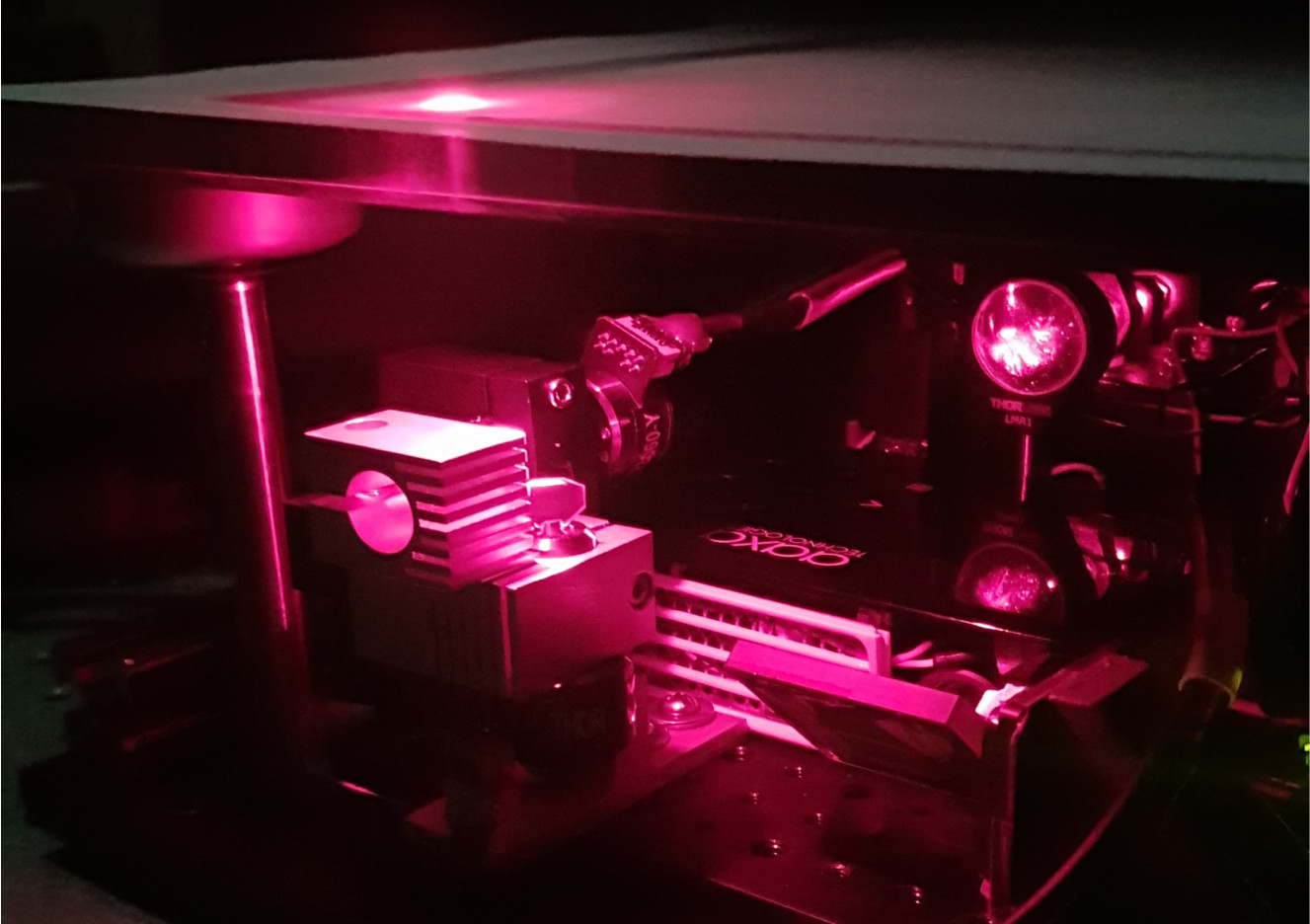
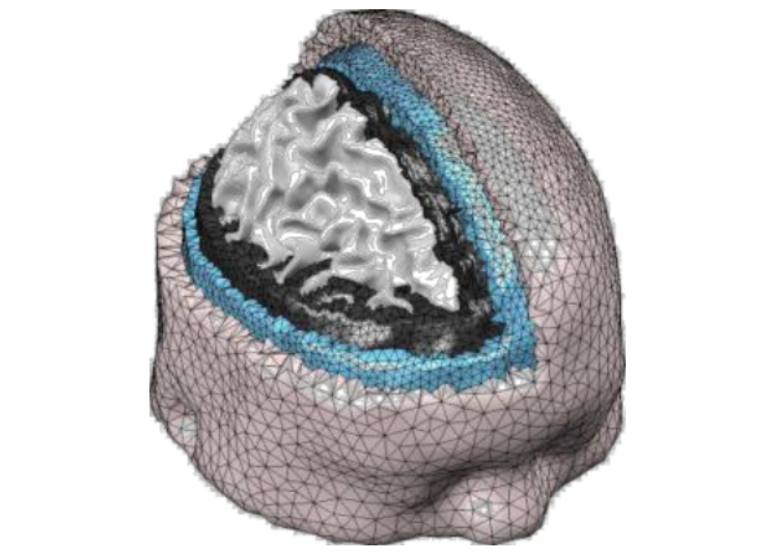
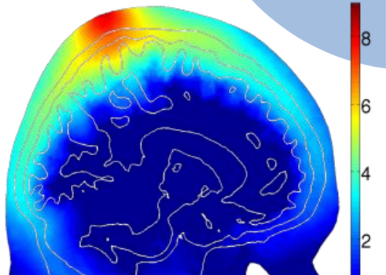
Biophotonics techniques play increasingly important roles in drug discovery, disease diagnosis, medical interventions as well as fundamental science research. The success of many biophotonics techniques is critically dependent on the ability to model complex interactions between photons and biological tissues accurately and efficiently. In 2009, Dr. Fang published one of the first papers using graphics processing units (GPUs) to accelerate 3D photon transport simulations. His code demonstrated a revolutionary 300-1000x acceleration and was considered “game-changing” by Dr. Simon Arridge (UCL), a pioneer in optical tomography. In 2010, Dr. Fang published a new mesh-based Monte Carlo algorithm. This paper became the most downloaded paper for Biomedical Optics Express in 5 consecutive months in 2013! So far, these works have attracted over 550 citations, 1,000 registered users and 15,000 downloads worldwide. Our open-source Monte Carlo simulation packages are actively used by many optical imaging research labs across the world. Our registration data also indicates that over 30 global companies utilize our software in their development, including Canon, Sony and Ricoh. In 2015, we were awarded an NIH R01 grant to continue leading this large user community and the development of novel massively parallel computational techniques.
-
 Translational Cancer Imaging
-
 Biomedical Optics
-
 Monte Carlo eXtreme
-
 Point-of-care Devices
-
 Multi-modal Imaging