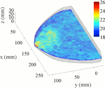
DigiBreast
 DigiBreast is a numerical breast phantom designed for 3D multi-physics simulations and validations of model-based image reconstruction algorithms for mammographically compressed breasts. The development of this phantom was described in Deng2015 with the original intent of testing a structure-prior guided image reconstruction algorithm for combined x-ray mammography and diffuse optical tomography (DOT) imaging. This digital breast phantom contains generic information such as 3D breast shapes and internal anatomical structures. We believe such breast phantom can address the needs for simulation-based validations for a wide range of model-based imaging modalities. Potential utilities of this digital phantom include, but not limited to, simulations of breast deformation, 2D and 3D x-ray breast imaging, and tomographic imaging of a compressed breast using tomographic optical, microwave, thermal and electrical impedance methods.
A unique aspect of this digital breast phantom is the inclusion of a realistic 3D glandularity map measured through a dual-energy x-ray mammography system, provided by Philips Healthcare. In comparison, conventional numerical breast phantoms represent various breast tissue constituents, i.e. the fibroglandular and adipose tissue, by piece-wise-constant regions (using a binary segmentation algorithm). Such representation removes the fine spatial details in the breast anatomical images, and results in loss of information. Statistical, or fuzzy segmentation methods avoid such information loss, and provide spatially-varying tissue volume fraction maps. In our previous works Fang2010, we have reported a joint x-ray/DOT image reconstruction algorithm utilizing a spatially varying tissue compositional model to improve DOT image resolution. This method was further characterized in Deng2015.
The DigiBreast phantom has limitations. While the breast shape is in 3D, the internal tissue compositional maps were derived from 2D x-ray measurements, thus, have an overall "cylindrical" shape along the sagittal direction. We, however, believe this approximation has negligible impact to most potential applications which deal with a mammographically compressed breast. This is because most of these methods utilizing a parallel-plate based measurement scheme and such scheme has an anisotropic spatial resolution - the horizontal/axial has significantly higher resolution than in the vertical/sagittal direction. Therefore, the focus in most of these imaging modalities are in the axial/horizontal view instead of the sagittal view. This limitation can be overcome in the future when 3D x-ray spectral imaging becomes available.
